DISCOVER THE DEEP VENOUS APPROACH:

A Structured Approach to Efficient Deep Venous Care
Deep venous care is often slowed by delayed diagnosis and fragmented referrals. The BD Deep Venous Approach structures the pathway from diagnosis to intervention to improve efficiency.
 Locked
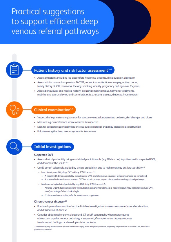
Practical suggestions PDF (3 Min Reading Time)

1. Diagnosis & Referral Pathway
Efficient referral pathways are critical to reducing delays in deep venous care. Studies show that Delays in diagnosing and fragmented referral systems can prolong diagnosis and treatment, impacting patient outcomes. BD Venous Solutions offer an efficiency-driven approach inspired by leading venous centers, supporting clinicians in optimizing referral pathways for deep venous disease.
More from Diagnosis & Referral Pathway
 Locked

Dr. Avgerinos (5:31 min)
 Locked
Dr. Baccellieri (4:28 min)
 Locked
Dr. Lichtenberg (3:42 min)
 Locked
Dr. Jalaie (4:28 min)
 Locked
Dr. Ortega (3:42 min)

2. Multidisciplinary Evaluation
Siloed decision-making leads to inconsistent treatment choices and delays, especially for patients who may require advanced interventions. ESVS guidance emphasizes involving vascular surgery, hematology, and interventional teams early to ensure timely and informed management.
BD Venous Solutions offers an efficiency approach inspired by leading venous centers, providing programs and solutions designed to streamline multidisciplinary collaboration.
Unlock to watch
Locked
The Power of Collaboration: Why Multidisciplinary Teams Matter in Venous Care
Dr. Baccellieri (4:28 min)
Locked
Driving Deep Venous Efficiency for Enhanced Patient Outcomes PDF (3 Min Reading Time)

3. Clinical Pathway & Intervention
Managing complex deep venous disease requires reliable, versatile solutions. Evidence-based guidelines recommend endovascular interventions such as stenting, thrombectomy, and PTA for restoring venous flow and preventing complications (ESVS Guidelines, 2022).
BD offers a comprehensive portfolio of stents, thrombectomy devices, and filters designed to support efficient, minimally invasive procedures — helping clinicians treat more patients effectively for longer.
Insights from the experts
Shaping venous care.
 Locked
 Locked
The Power of Collaboration: Why Multidisciplinary Teams Matter in Venous Care
Watch Video Locked
 Locked
 Locked
Risk Factors, Early Diagnosis, and Structured Approaches in Deep Venous Disease
Watch VideoEfficiency in action
Implementing efficiency in DVT/PTS preparation and stenting
Discover proven strategies for efficient DVT and PTS stenting, including anatomical insights, treatment algorithms, and classification systems that help optimize patient outcomes. Download the presentation to access case studies and expert tips designed to elevate your venous intervention practice.
Sign up to access the efficient approach to venous care
Stay in contact with us
Browse all upcoming venous events and access key details, dates, locations, and available content in one place.



Frequently Asked Questions
Deep veins are the main blood vessels that carry blood from the legs back to the heart. One-way venous valves and the calf muscle pump (the “peripheral heart”) keep blood flowing upward against gravity and prevent backflow. When valves are damaged or a vein is blocked, pressure rises in the leg and symptoms can develop.
Deep venous disease refers to chronic problems in the deep venous system caused by:
· Obstruction (residual clot, venous scarring, or compression—often iliac/iliofemoral), and/or
· Reflux (valves no longer close, causing backward flow).
Both raise venous pressure (venous hypertension), drive symptoms, and can lead to skin changes and venous ulcers.
Obstruction means a deep vein is narrowed or blocked—commonly after a DVT that did not fully resolve. It impairs outflow, elevates pressure, and can cause swelling, discomfort, venous claudication (effort-induced leg pain/cramping), and non-healing ulcers.
Reflux happens when damaged valves allow blood to fall backward in the deep veins. Persistent reflux sustains venous hypertension, worsening symptoms and skin damage over time.
DVT is a blood clot that forms in a deep vein, most often in the leg. It can partially or completely block blood flow. DVT matters because the clot can damage valves (leading to chronic symptoms) and break off, traveling to the lungs as a pulmonary embolism (PE).
Why DVT forms (Virchow’s triad):
· Stasis (slow flow): immobility, long travel, hospitalization, plaster casts.
· Hypercoagulability: cancer, pregnancy/postpartum, estrogen therapy, inherited thrombophilia.
· Endothelial injury: surgery, trauma, prior thrombosis, central venous catheters. [Ref 1, Ref 2]
Common risk factors: prior DVT/PE, recent surgery or trauma, active cancer, pregnancy, hormonal therapy, prolonged immobility, obesity, increasing age, smoking, and inherited predispositions.
PE occurs when a piece of a venous clot travels to the lungs, blocking blood flow and straining the right heart.
Symptoms: sudden shortness of breath, chest pain (worse with breathing), fast heart rate, coughing (sometimes with blood), fainting, or collapse.
Venous Thromboembolism (VTE) is the umbrella term for DVT and PE. Many PEs originate from leg DVTs. Without treatment, part of the clot can dislodge and embolize to the lungs.
PTS is a chronic condition months to years after DVT, caused by residual obstruction plus reflux.
Symptoms: leg ache/heaviness, swelling, tightness, cramps, itching; signs include skin discoloration, lipodermatosclerosis, and venous leg ulcers in severe cases.
· Annual VTE incidence in high-income settings is commonly reported around 1–2 per 1,000 people, increasing sharply with age.
· Global burden: VTE is among the most frequent cardiovascular conditions worldwide and a leading cause of cardiovascular mortality after myocardial infarction and stroke.
· Post-thrombotic syndrome (PTS) develops in a substantial minority after DVT; severe forms (with ulcers) occur in a smaller subset.
· Anticoagulation is the mainstay (DOACs, LMWH, or VKA when appropriate).
· Thrombolysis/thrombectomy may be considered in selected iliofemoral DVT to reduce clot burden or in high-risk PE with hemodynamic compromise.
· Compression started after acute pain/swelling allow, to help symptoms.
Foundational measures:
· Compression therapy to reduce venous hypertension and control symptoms.
· Lifestyle: leg elevation, calf-muscle activation/ambulation, weight management, smoking cessation, and skin care.
When obstruction is significant (especially iliac/iliofemoral):
· Endovascular venous stenting is commonly preferred when conservative therapy fails—especially with severe PTS, venous claudication, or non-healing venous leg ulcers.
· In combined obstruction + reflux, ESVS guidelines recommends treating obstruction first, which can alleviate reflux-related symptoms and clarify the need for further interventions.
When deep venous reflux persists despite optimal care:
· Surgical valve repair/reconstruction can be considered in specialized centers for select patients with significant, persistent symptoms.