Veterinary IV catheter placement: how to manage challenges
Peripheral intravenous catheters (PIVCs) are frequently placed in veterinary patients to administer fluids and medications.1 However, these vascular access devices (VADs) are associated with a risk of mechanical, inflammatory and infectious intravenous (IV) complications.1 IV catheter placement presents specific challenges for nonhuman patients.
Challenges for veterinary IV catheter placement
Anxiety and stress
A veterinary care setting can provoke anxiety in nonhuman patients because of smells, noise, separation from owners, unfamiliar environments and interactions with other patients.2 Feline patients are especially sensitive to their surroundings and may react to stress in ways that impede clinical evaluation and treatment.3
Risk of IV complications
PIVC complication rates tend to be lower in canine and feline patients than in humans.4 The overall PIVC complication rate in canine patients has been shown to be up to 20%.4 It is estimated at 35–50% in human patients.5
Veterinary patients with moderate to strong reactions to IV catheter insertion may affect device stability, leading to the possibility of mechanical IV complications.1 When there is more than one insertion attempt, IV complications have been shown to be more likely to occur in canine and feline patients.1
Damage to the endothelial layer during IV catheter placement may result in IV complications, such as phlebitis, extravasation or pain.6 If peripheral veins are catheterised multiple times, this may lead to venous depletion.4 Other IV complications reported in veterinary patients include catheter removal by the patient and line breakage, which is common in the intermediate care unit.4 This is why catheter securement is crucial in veterinary vascular access management.
Risk of exposure to zoonotic pathogens
As with human patients, blood can splash or spill from the IV catheter during insertion.7 Veterinary staff have reported significantly high needlestick injury rates (around 80%).8 Veterinarians carry an elevated risk of infection from zoonotic diseases.9 “Zoonotic” means they can be transmitted between humans and other vertebrate animals.10 The following factors should also be taken into account before placing an IV catheter in a veterinary patient.
Considerations in veterinary IV catheter placement
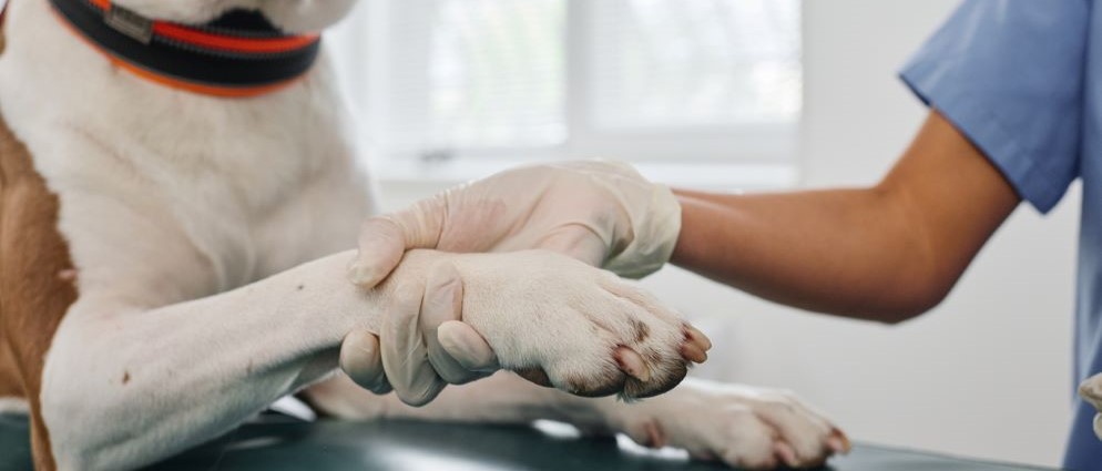
The most common peripheral insertion site for canine and feline patients is the cephalic vein.11 If the forelimbs are not available for IV catheter placement, then it may be necessary to place the IV catheter in the lateral saphenous vein in a hind limb.11 Before IV catheter placement, the veterinary patient should be comfortable and calm. It may be necessary to sedate the patient before insertion.4
IV catheter design has been shown to impact the risk of IV complications in humans.5 It has been demonstrated in human patients that rates of mechanical phlebitis vary based on the plastic composition and surface characteristics of IV catheters.5 Chhugani et al found that PIVCs made of polyurethane had lower rates of phlebitis in humans when compared with PIVCs made of polytetrafluoroethylene.12
Catheter hubs designed with an integrated septum have been shown to prevent blood leakage during insertion.7,13 Catheter gauge is an important consideration. Most canine patients can be placed with 20 G IV catheters.11 Larger canine patients can handle an 18 G.11 It is generally recommended that feline patients be placed with a 22 G.11 For smaller animals, 22 G or 27 G may be used.14
Schears et al reported lower rates of phlebitis in human patients whose IV catheters were secured with catheter stabilisation devices when compared to those secured with tape.15
10 practice recommendations to help optimise veterinary IV catheter placement
- Select a PIVC with a septum that enters the catheter hub to help control blood flow at insertion, connections and disconnections,7,13 eliminating the need for venous compression and reducing the risk of blood exposure. 13,†,*
- Use a PIVC made of material that softens in the vein to help minimise re-starts and reduce the risk of mechanical phlebitis.5,12,16,17,*
- Adapt the catheter gauge to the size of the animal.11,14
- Choose an appropriate insertion site based on the patient’s status and condition.11
- Remove all fur on the part of the limb where you’ll have the insertion site, catheter hub and connections.11
- Use aseptic technique to prepare the insertion site.18,*
- Ask your veterinary assistant to restraint the patient in the sternal recumbency position, while extending the limb where the IV catheter will be placed.11
- Have your veterinary assistant raise the vein by rolling it from medial to lateral with their thumb.11
- After insertion, secure the PIVC with a catheter stabilisation device.15,*
- Attach an extension set to help limit movement of the catheter hub.19,*
Find out more about veterinary vascular access management by discovering the Infuse 4 VetsTM website.


References
- Crisi PE, De Santis F, Aste G, et al. Inflammatory, Mechanical and Infectious Complications Associated with Peripheral Intravenous Catheters in Dogs and Cats: A Risk Factor Analysis. Vet Sci. 2022;9(3). doi:10.3390/vetsci9030118
- Lloyd JKF. Minimising Stress for Patients in the Veterinary Hospital: Why It Is Important and What Can Be Done about It. Vet Sci. 2017;4(2). doi:10.3390/vetsci4020022
- Carney HC, Little S, Brownlee-Tomasso D, et al. AAFP and ISFM feline-friendly nursing care guidelines. J Feline Med Surg. 2012;14(5):337-349. doi:10.1177/1098612X12445002
- Simpson SE, Zersen KM. Incidence and type of peripheral intravenous catheter complications documented in hospitalised dogs. J Small Anim Pract. 2023;64(3):130-135. doi:10.1111/jsap.13574
- Helm RE, Klausner JD, Klemperer JD, Flint LM, Huang E. Accepted but unacceptable: peripheral IV catheter failure. J Infus Nurs. 2015;38(3):189-203. doi:10.1097/NAN.0000000000000100
- Kuş B, Büyükyılmaz F. Effectiveness of vialon biomaterial versus teflon catheters for peripheral intravenous placement: A randomized clinical trial. Jpn J Nurs Sci. 2020;17(3):e12328. doi:10.1111/jjns.12328
- Haeseler G, Hildebrand M, Fritscher J. Efficacy and ease of use of an intravenous catheter designed to prevent blood leakage: a prospective observational trial. J Vasc Access. 2015;16(3):233-236. doi:10.5301/jva.5000334
- Mesquita JR, Sousa SIV, Vala H, Nascimento MSJ. The epidemiology of blood-contaminated needlestick injuries among veterinarians in Portugal. J Agromedicine. 2015;20(2):160-166. doi:10.1080/1059924X.2015.1010061
- Baker WS, Gray GC. A review of published reports regarding zoonotic pathogen infection in veterinarians. J Am Vet Med Assoc. 2009;234(10):1271-1278. doi:10.2460/javma.234.10.1271
- Habib I, Alshehhi Z. Zoonotic Disease Management and Infection Control Practices Among Veterinarians in the United Arab Emirates. Vet Sci. 2021;8(5). doi:10.3390/vetsci8050082
- Phillips H. Placing IV Catheters – Hints, Tips and Avoiding Common Mistakes for Vet Nurses. Published March 7, 2018. Accessed May 17, 2024. https://vetnurse.com.au/2018/03/07/placing-iv-catheters/
- Chhugani M, Mary James M, Thokchom S. A Randomized Controlled Trial to Assess the Effectiveness of VialonTM Cannula Versus Polytetrafluoroethylene (PTFE) Cannula in Terms of Indwelling Time and Complications in Patients Requiring Peripheral Intravenous Cannulation. Int J Sci Res IJSR. 2015;4(12):1075-1080.
- Onia R, Eshun-Wilson I, Arce C, et al. Evaluation of a new safety peripheral IV catheter designed to reduce mucocutaneous blood exposure. Curr Med Res Opin. 2011;27(7):1339-1346. doi:10.1185/03007995.2011.581275
- Hoareau G. Emergency care for kittens. Vet Focus Royal Canin. 2019;29(1). Accessed May 17, 2024. https://vetfocus.royalcanin.com/en/scientific/emergency-care-for-kittens
- Schears GJ. Summary of product trials for 10,164 patients: comparing an intravenous stabilizing device to tape. J Infus Nurs. 2006;29(4):225-231. doi:10.1097/00129804-200607000-00009
- Maki DG, Ringer M. Risk factors for infusion-related phlebitis with small peripheral venous catheters. A randomized controlled trial. Ann Intern Med. 1991;114(10):845-854. doi:10.7326/0003-4819-114-10-845
- Zhang L, Keogh S, Rickard CM. Reducing the risk of infection associated with vascular access devices through nanotechnology: a perspective. Int J Nanomedicine. 2013;8:4453-4466. doi:10.2147/IJN.S50312
- Nickel B, Gorski L, Kleidon T, et al. Infusion Therapy Standards of Practice, 9th Edition. J Infus Nurs. 2024;47(1S Suppl 1):S1-S285. doi:10.1097/NAN.0000000000000532
- French Society for Hospital Hygiene (SF2H). Prévention des infections liées aux cathéters périphériques vasculaires et sous-cutanés. Published May 2019. Accessed May 17, 2024. https://www.sf2h.net/publications/prevention-des-infections-liees-aux-catheters-peripheriques-vasculaires-et-sous-cutanes-mai-2019.html
This list of references to third-party peer-reviewed material and the sites they are hosted on are provided for your reference and convenience only, and do not imply any review or endorsement of the material or any association with their operators. The Third-Party References (and the Web sites to which they link) may contain information that is inaccurate, incomplete, or outdated. Your access and use of the Third Party Sites (and any Web sites to which they link) is solely at your own risk.
†Compared to a non-safety, non-blood control catheter.
*Recommendation for humans that could be applied to veterinary patients
BD-126487


